|
Experimental
Acute Experiments
IV. IABP in Acute
Myocardial ischemia
Summary – epicardial
mapping
In these experiments, we
attempted to quantitate: 1/ the effectiveness of intra-aortic ballon
pumping (IABP) in reducing severity and extent of myocardial ischemia,
2/ the persistence of induced changes, after the cessation of pumping,
3/ the effects of the duration of pumping, 4/ the effects of delaying
its application and, finally 5/ the effects of reperfusion. In dogs,
ligation of the left anterior coronary artery was followed by one hour
of observation of the natural progression of ischemia, then by the
IABP assistance and finally, additional one or 2 hours of observation,
to assess the persistence of the effects of pumping and additionally,
in some experiments, by reperfusion. Measurements were performed on
the ST segments and R and Q waves of the epicardial electrograms to
assess the severity (Σ ST – sum of ST segment elevations in mV) and
extent (NST – number of electrode sites with ST segment elevations) of
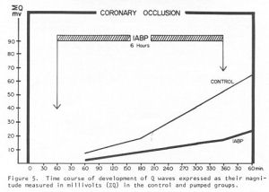
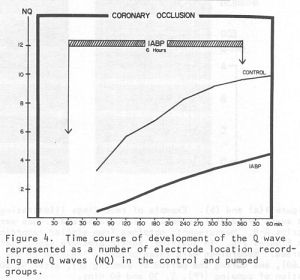
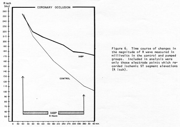
ischemia, and the R wave voltage loss and new Q wave development (Σ Q
and NQ), to assess the development of permanent damage and scarring.
The results were compared with the control groups.
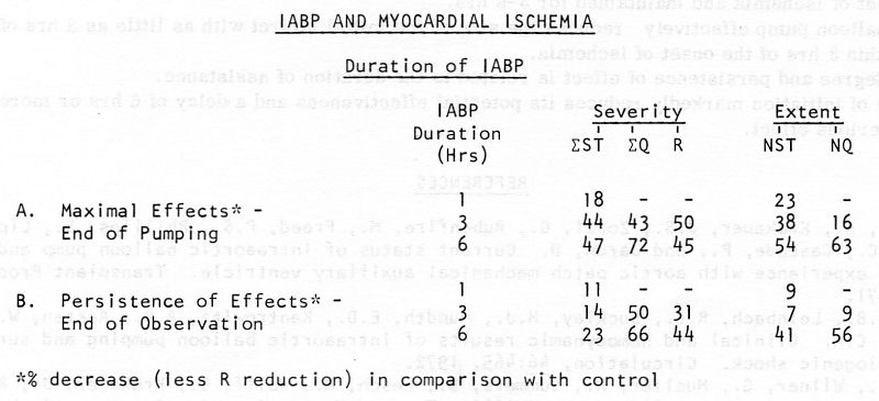
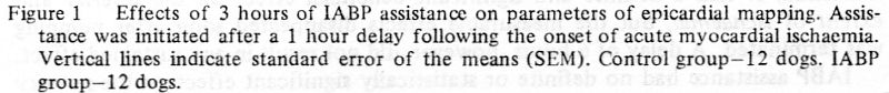
IABP – duration of pumping
The effects of varying
duration of pumping were evaluated in 3 groups of dogs, in which
the assistance
was initiated one hour after the onset of ischemia and continued for
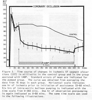
1 hour (12 dogs), 3 hours (12 dogs) or 6 hours (16 dogs). When
pumping was initiated one hour after the onset of ischemia and
continued for 1 hour - within 5 minutes, the severity of ischemia (Σ
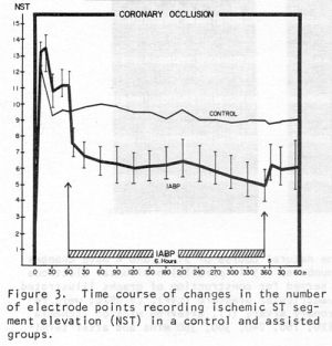
ST) was decreased by 15% and its extent (NST) by approximately 15%. At
the end of one hour of pumping Σ ST was decreased by 18% and
NST by 23%. The effects of pumping lasted in this group only as long
as pumping was continued and the measured parameters returned to their
pre-pumping levels within 5 minutes after cessation of pumping. In the
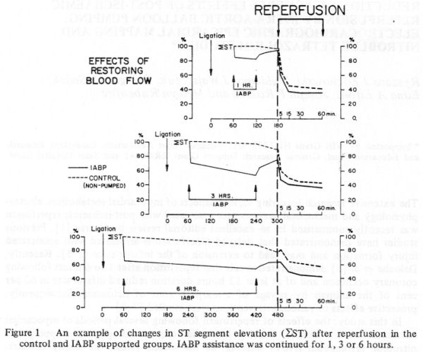
group with pumping continued for 3 hours, the severity of
ischemia (Σ ST) was decreased initially by 33% and its extent (NST)
by approximately 20% and their maximal reduction was by 44 and 38%.
The beneficial effect of assistance lasted longer, as the ST segment
elevations after cessation of pumping generally remained below their
pre-pumping levels during the period of post-pumping observation.
Similar changes were observed in regard to the extent of the ischemic
area. There was markedly less (40-50%) Q wave development and R wave
loss. When pumping was continued for 6 hours, the maximal
effects were a 47% reduction of Σ ST at the end of pumping, (in
comparison with the control group), but only a 23% reduction at the
end of an additional hour of observation (i.e. - partial recurrence of
the ischemic ST segment elevations).However, the extent of the
ischemic area (NST) was maximally reduced by 54% and it was still 41%
smaller (than in the control group) at one hour after termination of
pumping. There was 72% less Q wave development (Σ Q) at maximum
and still 66% less one hour later; the extent of the new Q wave zone
was 63% smaller at maximum and still 56% smaller at the end of
observation. There was 45% less R wave loss.
(Sedek, et al. IABP in acute
myocardial ischemia…)
1 +
1
+ 1 – (1 hour of ischemia + 1 hour
of IABP + 1 hour of reperfusion)
1 +
3
+ 1 - (1 hour of
ischemia + 3 hours of IABP + 1 hour of reperfusion)
1 +
6
+ 1
– (1 hour of ischemia + 6 hours of IABP + 1 hour of
reperfusion)
(Zochowski, et
al. Intra-aortic ballon pumping…)
 |
 |
 |
 |
|
|
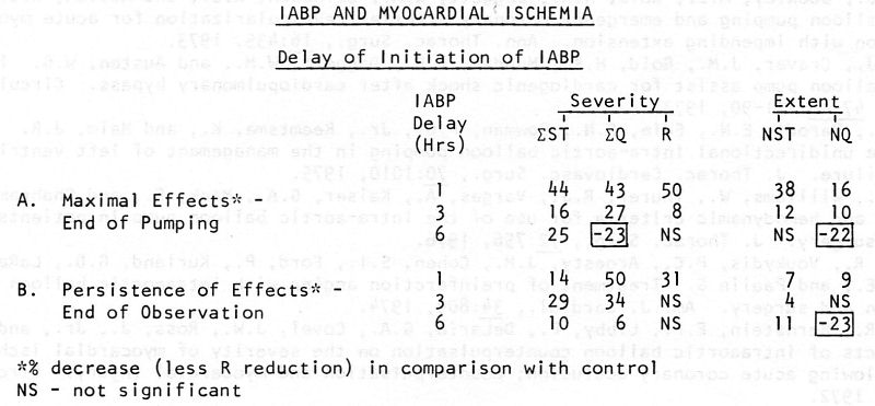
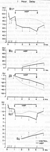
IABP – delay of pumping
The effects of varying delay in
initiation of pumping (with the same pumping duration of 3 hours)
was also studied in 3 groups of dogs – 1
hour (12 dogs), 3 hours (10 dogs) and 6 hours (10 dogs). The post-
pumping period of observation was extended to
two hours in the last 2 groups. The
results in a group with a 1 hour delay were described
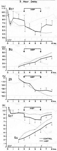
above. In a group with a 3 hours
delay, there was less reduction of ischemia (in comparison with
controls): Σ
ST was reduced by only 16% at the end of
pumping and by 29% at the end of observation. NST was
decreased by 12% and 4%, respectively.
Σ
Q was reduced by 27% and NQ by 10% at the end
of pumping and by 34% and NS (not
significant) at the end of observation. R wave loss was affected
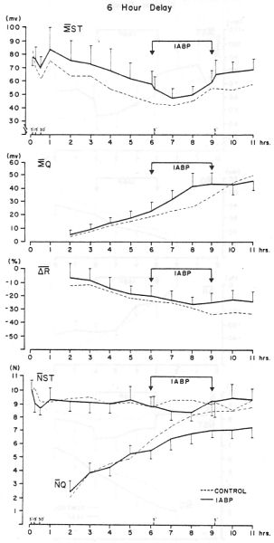
minimallyor not at all. 6 hour
delay in the initiation of pumping reduced the
Σ
ST by 25% at the end of the assistance and
byonly 10% at the end of observation and NST by 11%, only at the
end of observation. R waves were notaffected.
Σ
Q was 23% higher (in comparison with control)
at the end of pumping and NQ was 22% higher at the end of pumping
and 23% higher at the end of observation (accelerated development
of the Q waves!).
|
(Przybylski et al.
Intra-aortic balloon pumping…)
|
Figure 1 |
Figure 2 |
Figure 3 |
 |
 |
 |
| 1
+ 3 + 1 |
3
+ 3 + 2 |
6
+ 3 + 2 |


Publications:
1. Demonstration of lack of persistence of effectiveness of
intra-aortic balloon pumping of short duration in acute myocardial
ischemia. Sedek GS, Zochowski RJ, Wajszczuk WJ, Whitty AJ, Kiso
I, Freed PS, Moskowitz MS, Kantrowitz A, Rubenfire M. Trans Am Soc
Artif Intern Organs. 1975; 21: 555-65.
http://www.labmeeting.com/papers/author/wajszczuk-w
Experimental studies were carried out to quantitate the
effectiveness of intra-aortic balloon pumping (IABP) in reducing
severity and extent of myocardial ischemia and the persistence of
induced changes after cessation of pumping. Ligation of the anterior
descending coronary artery was followed by one hr of observation, IABP
for one hr (12 dogs) or 3 hrs (12 dogs) and an additional one hr of
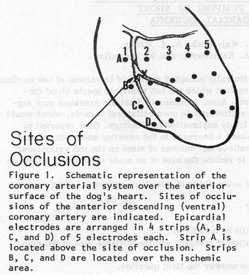
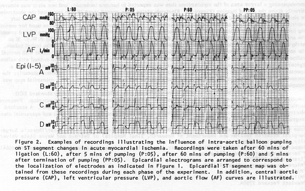
observation. Epicardial mapping utilizing 20 electrodes was used to
obtain the ST segment elevations (Sigma ST) and numbers of electrodes
showing ischemic ST changes (NST) in each group. Reductions of Sigma
ST of approximately 15% and 33% and reduction of NST of 15% and 20%
was observed in the one and 3 hr groups respectively, and persisted
throughout the period of pumping. Both parameters were noted to
increase within 5 min. after cessation of IABP in both groups. Sigma
ST frequently rose to almost pre-IABP values in the group pumped for
one hr. The group pumped for 3 hrs showed Sigma ST increase of
approximately 15% and NST increase of approximately 16%. Hemodynamic
measurements showed in both groups a mean systolic unloading of
approximately 10% and 10-20% mean diastolic augmentation. In
conclusion, IABP of short duration (1-3 hrs) early after the onset of
acute ischemia (one hr) induces a significant but transient decrease
in Sigma ST and NST, which reflects a reduction in myocardial ischemia.
Further study is required to evaluate the effectiveness of
intra-aortic balloon pumping, if initiated several hours after the
onset of ischemia, to reproduce the clinical reality of a patient with
an acute myocardial infarction
2.
Intra-aortic balloon pumping: Experimental relationships between
occlusivity and effectiveness. Wajszczuk, W.J., Sedek,
G.S., Whitty, A., Kiso, I., Freed. P.S., Moskowitz, M.S., Kantrowitz,
A. and Rubenfire, M. Med. Instr., 9:67, 1975.
3. Intra-aortic balloon
pumping in myocardial ischemia: The effect of pumping duration and
delay. Zochowski, RJ, Wajszczuk WJ, Przybylski
J, Sedek, GS, Kantrowitz A, Rubenfire M. Trans
Am Soc Artif Intern Organs. 1977,
23: 95-101.
4. Intra-aortic ballon
pumping during acute myocardial ischaemia – effects of delaying
initiation. Jacek Przybylski, Waldemar J Wajszczuk, Ryszard J
Zochowski, Mitchell S Moscowitz, Adrian Kantrowitz and Melvyn Rubefire.
Progress in Electrocardiology. Edited by Peter F. Macfarlane.
Pitman Medical. Publ. Co., Kent, England. 1979.
V. IABP and reperfusion
Summary
Publication:
1. Reduction of adverse effects of post-ischaemic reperfusion by
intra-aortic balloon pumping: electrocardiographic epicardial
mapping and nitroblue terazolium studies. Zochowski, Ryszard J.,
Wajszczuk, Waldemar J., Sedek, Grzegorz S., Elfont, Edna A.,
Roszka, Joseph P. and Rubenfire, Melvyn. Progress in
Electrocardiology, Edited by Peter W. Macfarlane. Pitman Medical
Publ. Co., Kent, England 1979, pp. 473-478.
2. Zochowski RJ, Wajszczuk W. Harmful effect of coronary
reperfusion after 5 and 8 hours of experimental myocardial infarct
in dogs. Protective role of intra-aortic balloon pumping].
Kardiologia Polska. 1981; 24(4):305-14.
Chronic Experiments
VI. IABP and chronic myocardial infarction
1. Summary – epicardial mapping and angiography
Effects of intra-aortic balloon pumping (IABP) on acute myocardial
ischemia (AMI) and chronic infarct
scar (CIS) induced by ligation of the anterior descending coronary
artery were studied in dogs. Epicardial
mapping with quantitation of ST, R and Q changes was correlated
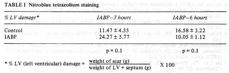
with nitroblue tetrazolium (NBT)
staining and angiography.
In acute phase experiments
(10 dogs) with 3 hours of IABP initiated with a delay of l hour after
the onset of acute ischemia, comparison with a control group (10
dogs) showed reduction of NST by 38% and NQ by 16% (extent of the
damaged myocardial zone). ΣST
(expression of the severity of ischemia) was reduced by 44%. There was
also 50% less R wave voltage reduction in the pumped group.
In chronic experiments, the extent of the CIS after 6 weeks was
reduced by 79% and 64% by Q and NBT mapping and there was 55% less R
voltage reduction. Postmortem angiography revealed development
of collaterals with ante- and retrograde filling of the distal
segments of the occluded vessels in pumped dogs Microangiography
revealed abundance of collaterals in pumped dogs.
In summary,
IABP is effective in permanently reducing the extent and severity of
ischemic myocardial damage. This effect is even more pronounced when
studied at 6 weeks. The ability of intra-aortic balloon pumping to
decrease the size of infarct scar in dogs has been
demonstrated.
Publication:
Experimental demonstration of the ability of intra-aortic balloon
pumping to reduce the infarct size. (Abstract, 26th Annual
Scientific Sessions of the American College of Cardiology).
Wajszczuk, W.J., Zochowski, R.J., Sedek, G., Elfont, E.E.,
Cascade, P., Roszka, J.P., Przybylski, J., Rubenfire, M. and
Kantrowitz, A. Am. J. Cardiol., 39: 259, 1977.
Summary - microangiography
Clinical evidence suggests that intraaortic balloon pumping
increases coronary blood flow to areas of ischemia in patients
with acute myocardial infarction. Microangiography was used to
determine the effects of balloon pumping on the development of
collateral vessels. Myocardial infarction was induced in dogs by
ligation of the ventral descending artery.
Stereo radiographs of the heart, before and after sectioning, were
obtained following injection of contrast medium (Micropaque) into
the coronary arteries. Vessels as small as 20 microns in diameter
could be visualized with this technique. Zones of avascularity
were clearly demonstrated in 3 of 4 control dogs, whereas 4 of 4
dogs supported by balloon pumping did not have avascular areas.
Collaterals were abundant in the pump group and were short,
straight, and generally under 100 microns in diameter.
Microangiography supports the theory that intra-aortic balloon
pumping following acute myocardial infarction increases
collateral flow to areas of ischemia and infarction.
Publication:
Microangiographic demonstration of increased blood flow to areas
of myocardial infarction during intraaortic balloon pumping. (Abstract,
43rd Annual Scientific Assembly of the American College of Chest
Physicians, Oct. 30 – Nov. 3, 1977). Cascade, P.N., Wajszczuk,
W.J., Kerin. N.Z. and Rubenfire, M. Chest 72, (3), 396, 1977
Summary – Myocardial ultrastructure, electron microscopy
When portions of cardiac muscle are deprived of blood flow,
infarct occurs and necrosis develops. The tissue immediately
surrounding the infarct is initially ischemic (Vikhert and
Cherpachenko, 1974). As healing proceeds, the infarcted area is
replaced by scar tissue but the fate of the ischemic zone is
unknown. The introduction of the IABP shortly after the initial
occlusion reduces the work load of the heart and increases
diastolic perfusion. This study concerns itself with the degree of
recovery of the initially ischemic myocardium surrounding the
established scar and the effect of the IABP on the degree of that
recovery.
Adult mongrel dogs of 25kg, were anesthetized
and a left thoracotomy was performed under sterile conditions. The
descending coronary artery was ligated and after a 1 hr
observation period, an IABP was introduced and pumping proceeded
for 3hr while electrophysiological recordings were made so that
epicardial ECG maps could be obtained. The chest was then closed.
Control animals underwent ligation but received no IABP. Six weeks
post-ligation, the chest was reopened and the heart mapped and
removed. Punch biopsies of normal, ischemic and scarred areas were
obtained immediately and fixed in cold 2% glutaraldehyde. The
entire heart was sliced and incubated in nitro-blue tetrazolium
(NBT) for identification of myocardial infarct (Nachlas and
Shnitka, 1963). The fixed tissue was post-fixed in 1 % OSO4 and
processed by routine methods for electron microscopy.
Comparison of maps of scarred and normal myocardium
prepared from NBT incubated heart slices 6 weeks post-ligation and
epicardial EGG maps of anoxic, ischemic and normal areas showed that
the myocardium surrounding the scar at 6 weeks was originally
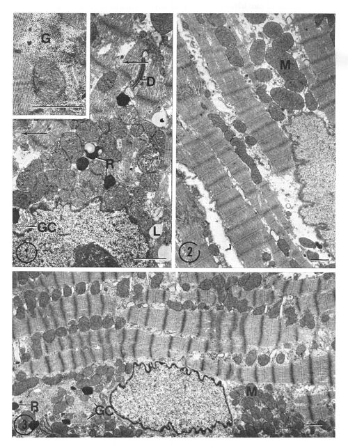
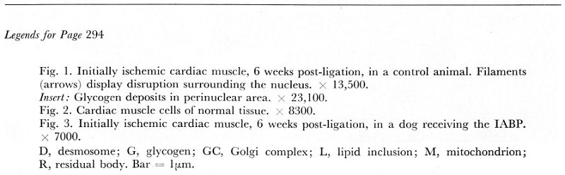
ischemic. Electron microscopic examination of this tissue in
control animals (Fig. 1), revealed a greater number
of intracellular glycogen deposits, perinuclear lipid inclusions and
residual bodies than seen in normal myocardium of the same animal
(Fig. 2). Although present, these changes were less pronounced in
pumped animals (Fig. 3). Thick and thin filaments near the nuclei of
cells of control animals showed occasional disruption and
disorganization as did those adjacent to intercalated discs. Cells
from pumped dogs did not display these alterations to the same degree.
We
have demonstrated that ultrastructural changes are present in
initially ischemic myocardium 6 weeks post-ligation of a coronary
artery. The extent of these changes would indicate that these cells
have not recovered normal function. The use of the IABP for 3 hrs
after a 1 hr delay appears to lessen the amount of persisting
morphological damage seen in initially ischemic tissue.
Publication:
Modification of the ultrastructure of myocardium adjacent to
chronically infarcted areas by the intra-aortic ballon pump (IABP).
Roszka, Joseph P., Elfont, Edna A., Kobernick, Sidney D.,
Zochowski, R.J. and Wajszczuk, W.J. Micron,1976, vol 7:
293-295, Pergamon Press, Printed in Great Britain.
Conclusions:
Acute experiments:
-
In experiments on anesthetized dogs, the balloon pump effectively
reduces the severity and extent of acute ischemia, when applied within
1-3 hours of the onset of ischemia and maintained for 3-6 hours.
IABP of short duration (1-3 hours) early after
the onset of acute ischemia (with a one hour delay) induced
significant but transient decrease in ΣST
and NST, which reflects a local reduction of the severity and extent
of ischemia; the effects tended to disappear (or markedly diminish)
shortly after termination of assistance.
The balloon pump effectively reduces the size
of the initial infarct with as little as 3 hours of assistance, when
applied within 3 hours of the onset of ischemia.
Three hours of pumping (initiated with a one
hour delay) appeared to decrease (or delay?) the Q wave development
and decrease (or delay?) the R wave loss.
The degree and persistence of its effects is
related to the duration of the assistance.
Six hours of pumping (initiated with a one hour delay) reduced by over
50% the extent of the ischemic zone, R wave
loss and Q wave development.
Delay of initiation markedly reduces its potential
effectiveness and a delay of 6 hours or more might result in a
deleterious effect – see comments below, regarding the chronic
experiments.
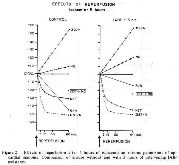
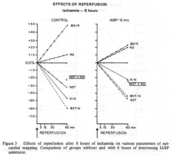
Reperfusion of the ischemic area after 5 and 8 hours of ischemia lead
to the acceleration of the Q wave development (“reperfusion injury”?)
in the control group, which was significantly diminished in the group
with balloon pumping.
Chronic experiments:
-
Re-evaluation in chronic experiments, after six weeks of recovery,
showed very marked beneficial
long-term effect of pumping:
The extent of the chronic infarct scar after 6 weeks was reduced by
79% by Q wave and 64% by NBT mapping and there was 55% less R voltage
reduction (in a group assisted by IABP for 3 hours after a 1 hour
delay).
Postmortem angiography revealed development of
collaterals with ante- and retrograde filling of the distal segments
of the occluded vessels in pumped dogs. Microangiography revealed
abundance of collaterals in pumped dogs.
Electron microscopy study study, in correlation
with NBT and electrocardiographic mapping, revealed significant
preservation of the ultrastructure in the initially ischemic
myocardium.
The response to IABP during the acute phase of
ischemia and infarction may not be used to accurately
predict the
long-term beneficial effects of pumping. (There was marked
discrepancy between the findings in the acute phase of the
experiments and those observed after 6 weeks).
|
)
)
)
)
)
)
)
)
)
)
)
)
)
)
)
)
)
)