|
Summary
Terminology used in clinical
electrocardiography includes terms: the “baseline” (or the T-P
interval) – which is accepted to be stable, (or made so by the type of
amplifiers used in the clinical recorders), of zero voltage, and then
the J- point and ST-segments shifts, (in relation to this stabilazed
baseline), which correlate well with various types of clinical
pathology and have a definite clinical significance. On the other
hand, certain types of amplifiers used in the experimental work in
laboratories, may introduce “drifting of the baseline” (requiring
constant correcting to allow successful recording), which is caused by
the “polarization effect” currents which develop at the interface
between the metal components of the recording electrodes and the live
tissue, body fluids and electrolyte content and concentration. We
became interested in these phenomena and decided to try to determine,
if there exist a “true” baseline shifts – in relation to a stable D.C.
(direct current) zero voltage baseline – and what are in reality the
“true” J-point and ST-segment shifts recorded in clinical
electrocardiography.
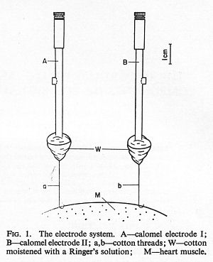
Pilot experiments were carried
out on the exposed hearts of anesthetized rabbits and dogs using
laboratory type calomel electrodes and D.C. amplifiers. Bipolar
epicardial leads were recorded. The same type of electrode was used as
a distant “grounding” electrode. Interventions included local
application of potassium and sodium solutions and induction of local
myocardial ischemia by ligation of small coronary artery branches.
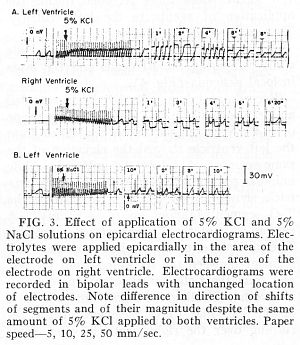
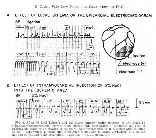
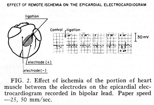
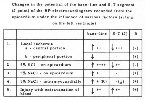
True baseline shifts were
observed, in both directions (positive or negative), alone or with the
J-point and ST-segment shifts, in opposite direction. They were most
pronounced after local application od potassium solution or severe
local ischemia. Examples of recordings are shown below and
observations are summarized in the Table. No attempt was made in these
limited series of experiments to elucidate the mechanism for creating
these voltage differentials between the baseline (T-P segment), the ST
segment and the zero D.C. baseline. It remains to be determined, if
this type of differentiation provides any new type of information,
carries any clinical significance or could be correlated with the type
or degree of clinical pathology.
1.
A Method for Experimental Recording of the D.C. and
Very Low Frequency Components of the Electrocardiogram. Waldemar J.
Wajszczuk and Józef K. Cywinski. Engineering in Medicine and
Biology, Proceedings of the 19th Annual Conference,
San Francisco, CA, 17 November, 1966, p.
245.
2.
The recording of d.c. and very low frequency
components of ECG signals from epicardial leads.
Józef K. Cywiński and
Waldemar J.
Wajszczuk
Medical and Biological Engineering and
Computing,
Volume 4, Number 2 / March, 1966
http://springerlink.com/content/?k=Wajszczuk;
https://commerce.metapress.com/content/58087405p5370075/resource-secured/?target=fulltext.pdf&sid=nbw12255rarcz3jdln4y4h55&sh=springerlink.com;
(https://springerlink3.metapress.com/content/58087405p5370075/resource-secured/?target=fulltext.pdf&sid=fezt2z45iyr3d355cgdmpq55&sh=www.springerlink.com)
http://www.springerlink.com/content/58087405p5370075/
Abstract.
By means of the calomel
electrode technique described and the use of
d.c.
amplifiers it has been possible to measure and record the very low
frequency and d.c. components of ECG signals.
These signals can be recorded by means of a commercial
electrocardiograph with 50 mV
d.c. input terminals, connected to
a special preamplifier. Both very low frequency and
d.c. components of signals from
the epicardium carry information about the heart muscle conditions.
These conditions depend, among others, on the electrolyte composition
and concentration changes in a zone between two exploring electrodes
on the heart muscle.
http://www.ncbi.nlm.nih.gov/sites/entrez
3.
Observations on D. C.
and Very Low Frequency Components of the Electrocardiogram.
Waldemar J. Wajszczuk
and Józef K. Cywinski. Exp. Biol. Med. 123(1): 42—47; Volume 123
Number 1, October 1966
http://ebm.rsmjournals.com/content/vol123/issue1/ |
)
)
)
)
)